
Improving Accuracy in Biological Research
AI 'Ramanomics' Enables Dye-Free Living Cell Imaging
University at Buffalo researchers use AI and Raman spectroscopy to identify organelles without invasive dyes.
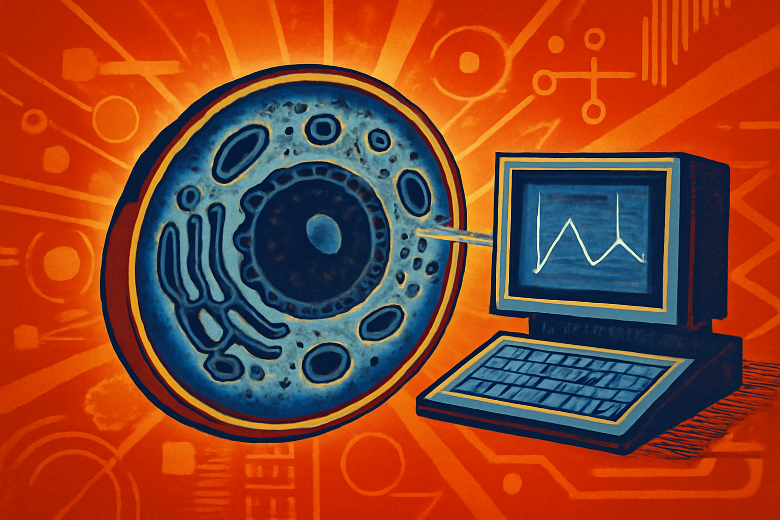
A digital rendering of a living cell with distinct organelles highlighted by light patterns, representing AI-driven Ramanomics imaging in a laboratory context.
Photo: Kronos Digital News
Researchers at the University at Buffalo have developed a new method called "Ramanomics" to study living cells [1]. This breakthrough combines artificial intelligence with Raman spectroscopy to identify cellular organelles using their unique biochemical fingerprints [1]. By using light instead of chemical markers, scientists can observe cell behavior without interference [1].
Traditional methods often rely on fluorescent dyes, which can disrupt natural biological processes [1]. These dyes may also limit the accuracy of measurements in long-term research [1]. The Ramanomics approach offers a non-invasive alternative that preserves the integrity of the living specimen [1].
Editorial notes
Transparency note
AI assisted drafting. Human edited and reviewed.
- AI assisted
- Yes
- Human review
- Yes
- Last updated
Risk assessment
Risk level set to high because the story relies on a single source domain (University at Buffalo).
Sources
Related stories
View all


Topics
About the author
Kronos Digital News Desk covers improving accuracy in biological research and editorial analysis for Kronos Digital News.